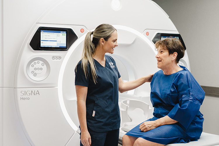
fbpx

Magnetic Resonance Imaging (MRI)

With its remarkable ability to capture detailed images in a non-invasive and pain-free manner, MRI technology plays a pivotal role in providing accurate diagnoses and personalised treatment options, ensuring the highest quality of care for our patients.

Using MRI, South Coast Radiology’s specialist radiologists are able to obtain highly detailed and comprehensive images of internal structures and processes, providing invaluable information to aid in the diagnosis and treatment planning of various medical conditions.

  • What is Magnetic Resonance Imaging?

    Magnetic Resonance Imaging (MRI) uses an exceptionally strong magnet, radio frequency waves and a sophisticated computer to generate detailed high-quality images of structures and processes within your body. MRI provides helpful information to your doctor which aids the diagnosis of your condition and planning your treatment options.

    This innovative imaging technique allows us to capture images from multiple perspectives, providing a comprehensive view of the target area within the body. Whether it’s visualising the intricate network of blood vessels, evaluating the integrity of the musculoskeletal system, or exploring the complexities of the brain and nervous system, MRI offers unparalleled insights into the inner workings of the human body.

    The body or body part is placed within a magnetic coil that detects shifting signals, which are encoded by the magnetic field gradients. Finally, powerful computers process the signal to form images of the area of interest. These high-quality images provide information to your doctor about structures and processes within your body. This helps your doctor diagnose your condition and plan your treatment options.

    At South Coast Radiology, we have two fully Medicare-eligible MRI services at John Flynn Private Hospital and our Benowa clinic on the Gold Coast, in addition to two partially eligible MRI locations at Smith Street and Robina. We accept MRI referrals from Medical Specialists, GPs and Physiotherapists. GPs have access to a limited number of MRI examinations for both adult and paediatric patients. Medical Specialists have access to the majority of MRI examinations, however, not all MRI examinations attract a Medicare rebate.

    Our MRI bookings team can discuss your options regarding whether a Medicare rebate is available for your scan, at the time of your booking.

  • BEFORE AN MRI SCAN

    Because a strong magnet is used in Magnetic Resonance Imaging scans, patients with certain ferromagnetic or electronic implanted devices or prostheses may not be able to undergo an MRI procedure. When a patient arrives, he or she will be asked to answer a series of safety questions. Patients with cardiac pacemakers, metal heart valves, some ear implants, certain brain aneurysm clips and various other medically implanted devices may not be able to have an MRI. This is because the metal or electronics will respond to the strong magnetic field.

    We may require further information on certain devices to determine their compatibility in the MRI environment. Also, people with metallic foreign bodies in their eyes, through grinding or welding, may be excluded from MRI. An X-ray of the eyes may be required to clear the eyes of any metal fragments before you are cleared to undergo an MRI.

    Patients having a scan of the abdomen or pelvis may need to fast before the appointment, in order to avoid bowel and stomach movement. Our staff will advise if a patient needs to fast, and for how long they will need to do so when the booking is made.

    Patients who suffer from claustrophobia might not feel comfortable laying within the magnet for the required time. Patients should advise our staff prior when booking an appointment if they are claustrophobic so that we can discuss options.

  • DURING AN MRI SCAN

    The MRI machine combines an open-ended magnet with a padded bed, which moves through the centre. The body region of interest is also placed within a device called a ‘receiver coil’. Throughout the scan, the radiographer will maintain visual and voice contact with the patient. The switching of the magnetic gradients creates a loud thumping sound, so the patient will be given hearing protection, which must be worn during the scan. Different sites may also be able to offer music during your scan. Discuss this with the radiographer on the day to see if this is an option for you.

    You will be required to remain motionless for the entirety of the scan to obtain high-quality images. If you believe this could be a concern please notify our friendly booking staff when you make your appointment.

    Sometimes, a small dose of gadolinium-based contrast is injected through a vein in your arm. This is done to temporarily alter the magnetic properties of the body tissue and enhance anatomical detail. Our radiologist will determine whether an injection is required at the time of the scan.

  • WHAT HAPPENS AFTER AN MRI?

    MRI is painless and you will not feel any adverse after-effects, so you are able to resume normal activity straight away.

    One of our radiologists will interpret the scan and provide your referring doctor with a comprehensive report about the findings. The results of your scan will be sent directly to your referring doctor, and it is very important you book a timely follow-up appointment to discuss your results.

  • MRI PATIENT SAFETY

    MRI (Magnetic Resonance Imaging) is generally considered a safe and non-invasive diagnostic tool. However, there are certain safety considerations that patients should be aware of before undergoing an MRI.

    Patients with cardiac pacemakers, metal heart valves, some ear implants, certain brain aneurysm clips and various other medically implanted devices may not be able to have an MRI because the metal or electronics will respond adversely to the strong magnetic field. We may require further information on certain devices to determine their compatibility in the MRI environment. Also, people with metallic foreign bodies in their eyes, through grinding or welding, may be excluded from MRI. An X-ray of the eyes may be required to clear the eyes of any metal fragments.

    Patients should also avoid wearing any metal objects, such as jewellery or hair clips, during an MRI, as these can be pulled by the magnetic field and cause injury.

    During the MRI, the patient will need to lie still for an extended period of time, so it’s important to inform the technician if you have any conditions that may make it difficult to do so, such as claustrophobia.

    No X-rays or ionising radiation is used during an MRI. Additionally, the magnetic field and radiofrequency waves produced by Magnetic Resonance Imaging machines have not been shown to cause any long-term effects on the patient. In some cases, contrast agents may be used to enhance the images. These contrast agents are generally safe but may cause allergic reactions in some patients.

    Overall, MRI is a very safe diagnostic tool, but it’s important for patients to inform their doctor and technician of any potential safety concerns or medical conditions before undergoing the procedure.

Find out more about the specialist MRI services offered by South Coast Radiology

To find out more about a service, please click its name below.
Please note; not all services are listed below, and not all services are available at every site.

Breast MRI Scans

On average, 50 people will be diagnosed with breast cancer every day. Although the number of women and men being diagnosed in Australia is increasing, the number of deaths from breast cancer is decreasing. Breast MRI is an extremely sensitive, non-invasive, medical imaging examination used for the detection of breast cancer and other breast abnormalities. It can also be used to examine the extent of breast cancer after a diagnosis has been made following a mammogram, ultrasound and/or biopsy. The quality of images produced by MRI of breast tissue is outstanding.

MRI makes it easy to take additional images of the muscle and chest wall around your breast to provide a more comprehensive diagnosis.

Breast MRI can be beneficial in detecting breast cancer in high-risk women with a strong family history or a past history of breast cancer, or who are carrying a genetic predisposition to breast cancer.

For examining breast implants, mammography and ultrasound may not penetrate saline or silicone well enough to view the implants or the surrounding breast tissue. In contrast, MRI may offer clearer images of both implants and breast tissue. This makes MRI an excellent imaging modality to assess breast implant integrity and detects small ruptures or leaks.

Before a Breast MRI

You will require a referral from your doctor prior to making an appointment for a breast MRI. This exam is to be booked between days 6-16 of your menstrual cycle (if you still have cycles). Do not be concerned if you have an irregular cycle, as we will still make an appointment that best fits your current cycle.

On arrival to your appointment, our MRI technologist will explain the examination in detail and check your contraindication questionnaire. Once this has been checked by the MRI technologist that you are safe to have an MRI scan, they will prepare you for the scan by inserting an IV cannula for the MRI contrast to be injected during this test. You will also be changed into a patient gown prior to your scan.

For most breast MRI scans, an injection of a gadolinium-based contrast solution into your arm vein will be required. This is to enhance the detail of the images produced. MRI scans to assess implant rupture will not require this injection.

During a Breast MRI

The scans are taken as you lay on your stomach, and your chest will move into the centre of the MRI scanner. It is very important that you remain perfectly still while the images are being acquired and breathe gently to minimise any motion or movement of the breasts.

After an initial series of scans, the contrast material will be injected through the IV cannula. It is normal to feel a sense of coolness and a flushing sensation for a minute or two when the contrast is been injected. Additional series of images will be taken during or following the injection.

It will take approximately 30 to 45 minutes to complete an MRI of your breasts.

After a Breast MRI

There are no known risks from having a breast MRI. After the MRI examination, you should be able to resume your normal activities immediately.
A radiologist specialising in breast imaging will interpret your MRI scans and provide a comprehensive report on the findings to your doctor.

About Prostate MRI Scans

Prostate MRI is a non-invasive medical imaging examination that can detect prostate cancer and provide information about other prostate concerns.

Prostate cancer causes over 3000 deaths in Australian men each year. This makes prostate cancer the fourth main cause of death in Australian males. The risk of prostate cancer increases with age and family history. By the age of 85, approximately 1 in 5 Australian males will be diagnosed with prostate cancer.

If both a prostate-specific antigen (PSA) blood test and digital rectal examination return abnormal results, your doctor may request further tests or refer you to a specialist urologist for a diagnosis.

Until recently, prostate cancer was difficult to image and detect. MRI has emerged as the imaging technique of choice for detecting prostate cancer and provides important information for determining treatment options. MRI of soft tissue and organs (such as the prostate) are clearer and more detailed than other imaging methods. MRI enables early diagnosis of prostate cancer as well as an accurate evaluation of the tumour’s extent.

Why are MRI scans used to detect prostate cancer?

MRI can determine if the cancer is entirely inside the prostate gland or has spread to nearby or distant organs. For men with an intermediate or high probability of cancer spread outside the prostate, MRI improves the accuracy of information, allowing your doctor to determine a more appropriate treatment plan. MRI also provides important information to surgeons. Understanding the extent of cancer in your prostate helps your surgeon to preserve delicate arteries and nerves that are essential for sexual function and bladder control.

Furthermore, prostate MRI is used to evaluate other prostate problems such as infection or abscess, prostate enlargement, congenital abnormalities or complications after pelvic surgery.

Before a Prostate MRI

Before making your appointment, you will require a referral for a Prostate MRI from your Urology or Oncology specialist.  If your Prostate MRI referral meets Medicare criteria it will be bulk billed. Please speak with our dedicated MRI bookings agents to ask about prices, locations and services.

To achieve a successful MRI prostate scan, it is important that your bowel is empty for the procedure. You will be given 2 Durolax tablets to take the night before the procedure. These tablets should cause evacuation of your bowel early in the morning on the day of your procedure. We ask that you do not have an ejaculation for 48 hours prior to the scan. You must have nothing to eat, drink, smoke or chew for 6 hours prior to the appointment.

On arrival to your appointment, our MRI technologist will explain the examination in detail and check your contraindication questionnaire. Once this has been checked by the MRI technologist that you are safe to have an MRI scan, they will prepare you for the scan by inserting an IV cannula for the MRI contrast to be injected during this test.  You will also be changed into a patient gown.

An intramuscular injection will be administered 30 mins prior to the scan. The purpose of this medication is to relax the bowel to minimise motion which can interfere with the images.

During a Prostate MRI

During the MRI scan, you will be positioned on your back on the MRI bed and your pelvis will move into the centre of the MRI scanner.  It is very important that you remain perfectly still while the images are being acquired. If there is movement during the scan, the scan may not be of diagnostic quality.

After an initial series of scans, the contrast material will be injected through the IV cannula. It is normal to feel a sensation of coolness and a flushing sensation for a minute or two when the contrast is been injected. Additional series of images will be taken during or following the injection.

The scan will take approximately 30mins.  Please be aware you will be in the department for about 1 ½ hours.

After a Prostate MRI

There are no known risks or side effects from having a Prostate MRI.  After the MRI examination, you should be able to resume your normal activities immediately. A radiologist specialising in prostate imaging will interpret your MRI scans and provide a comprehensive report on the findings to your doctor. It is very important you book a timely follow-up appointment to discuss your results.

Cardiac MRI is a non-invasive technique used to assess the function and structure of the cardiovascular system. Utilising Magnetic Resonance Imaging technology, South Coast Radiology can assess myocardial viability, assess cardiomyopathy, measure ventricular volumes, investigate suspected myocarditis, characterise myocardial masses, and quantify blood flow across heart valves.

The scan will take 45-60 minutes to complete, and no special preparation is required. Before their procedure, patients will be given an MRI safety questionnaire to complete. During the procedure, ECG dots and leads will be placed onto the patient’s chest to monitor the heart during the scan, and a cannula will be inserted into the patient’s arm so a contrast agent can be given during the MRI. A lightweight blanket camera will be placed on the patient’s chest to acquire MRI images, and the patient will be required to hold their breath multiple times during the scan.

This form of medical imaging is tailored to the individual patient using a variety of methods and is able to overcome various limitations to acquire high-quality cardiac images.

World Class MRI Technology

South Coast Radiology has installed Australia’s first GE 3T MRI machine equipped with Air Recon DL Technology.

This first MRI AIR Tech delivers consistently superior image quality in a significantly reduced scan time. Meaning, the patient spends less time on the bed without any compromise on image quality.

MRI Scans on the Gold Coast

If you are looking to make an appointment for a Magnetic Resonance Imaging (MRI) scan, be sure to book online or call our dedicated bookings line by clicking below!