Our Services
Cardiac Imaging
At South Coast Radiology, our Cardiac Imaging services provide a detailed view of how your heart and blood vessels are functioning.
Using advanced imaging technology – including CT Coronary Angiography (CTCA), Echocardiography, Cardiac MRI, and Nuclear Cardiology (Myocardial Perfusion), our experienced radiologists and cardiac specialists deliver precise and timely information to help diagnose, manage and monitor a wide range of heart conditions.
We work closely with your referring doctor or cardiologist to ensure every test is tailored to your clinical needs, offering accuracy, comfort, and care you can trust.
What is Cardiac Imaging?
Cardiac imaging refers to a group of non-invasive tests that capture detailed images of your heart’s structure and function.
These tests help your doctors assess:
- The size, shape, and motion of the heart
- The condition of heart valves and chambers
- Blood flow and oxygen supply to the heart muscle
- The presence of blockages, plaque, or calcium in the coronary arteries
Our Cardiac Imaging Services:
- Echocardiography (Echo): Ultrasound imaging of the heart
- CT Coronary Angiography (CTCA): A detailed CT scan to view coronary arteries
- Calcium Scoring: Measures calcium build-up in arteries to assess future risk
- Myocardial Perfusion (Nuclear Cardiology): Evaluates blood flow to the heart muscle
- Cardiac MRI: Provides high-resolution images of the heart’s structure, motion, and tissue composition.
Why might I need cardiac imaging?
Cardiac imaging plays a vital role in identifying problems early — often before symptoms become serious.
Your doctor may refer you for cardiac imaging to:
- Investigate chest pain, palpitations or shortness of breath
- Detect or monitor heart disease
- Assess heart function after a heart attack or cardiac surgery
- Evaluate valve disorders or congenital heart conditions
- Plan and monitor ongoing treatment or prevention strategies
Which test is right for me?
Your cardiologist or GP will recommend the most appropriate test based on your symptoms, medical history, and risk factors.
Echocardiogram
CT Coronary Angiography
Calcium Score
Myocardial Perfusion
CARDIAC MRI
About Your Test

Before your appointment
Preparation varies depending on your test type. Our team will confirm your instructions when booking.
General guidelines:
- Avoid caffeine and smoking for 24 hours before your scan, as these affect heart rate.
- Avoid heavy exercise on the day of your scan.
- Wear comfortable, loose-fitting clothing and remove jewellery or metal objects near your chest.
- Bring your referral, Medicare card, and any previous imaging results.
- If you take heart medication, our team will advise if any adjustments are needed before your test.
- For CTCA or Calcium Scoring, you may be asked to fast for 2–3 hours prior.
- For CTCA scans, you may receive medication (such as a beta-blocker) to help slow your heart rate for clearer imaging.

On the day
Your experience will depend on which test you require.
Echocardiogram: You’ll lie on an examination bed while a cardiac sonographer moves a small ultrasound probe across your chest using warm gel. The test is painless and takes 20–30 minutes.
CTCA/Calcium Score: You’ll lie on a CT scanner table with ECG leads attached to monitor your heartbeat. You may feel a warm sensation if contrast dye is injected — this is normal and passes quickly. The scan itself takes less than 10 minutes.
Myocardial Perfusion Scan: A small amount of radioactive tracer highlights blood flow to your heart muscle. Two sets of images are taken — one at rest, one after gentle stress or medication — to show how well your heart is supplied with blood.
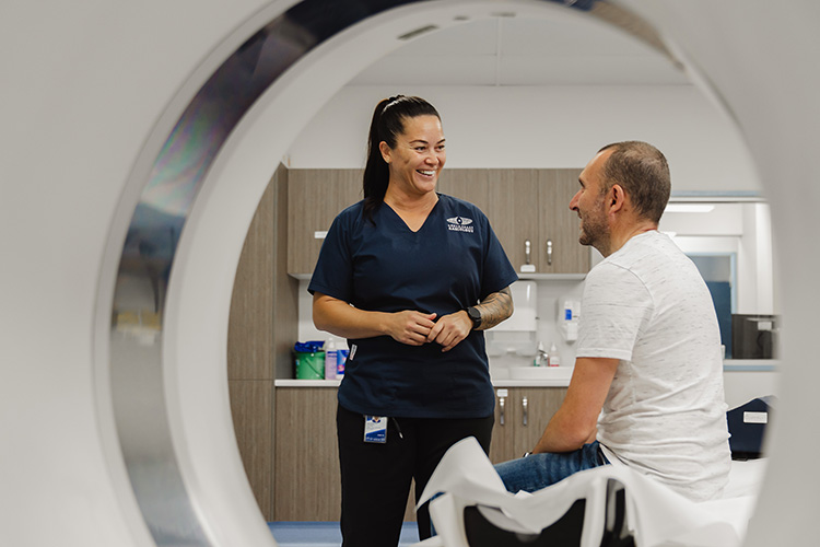
Cardiac MRI: You’ll lie inside a wide MRI scanner while detailed images are taken of your heart. You may be asked to hold your breath for short periods. The scan is painless and typically takes 30–60 minutes.

After your appointment
- You can usually resume normal activities immediately.
- If you received contrast or tracer, we recommend drinking plenty of water for the next few hours.
- Our specialist radiologists will interpret your results and send a comprehensive report to your referring doctor.
- Your referring doctor or cardiologist will review your report and discuss the findings with you.
Depending on your results, they may recommend lifestyle changes, further testing, or treatment options.
Our role is to ensure your doctor has accurate, detailed information to support your care and ongoing heart health
Frequently Asked Questions
Yes. These tests are non-invasive and use either ultrasound or low-dose radiation. For nuclear scans, the radioactive tracer is safe and leaves your body naturally within 24 hours.
Most tests take 15–45 minutes, depending on the study type. Myocardial perfusion scans may take up to 2–3 hours including rest and stress phases.
Yes. A referral from your GP or cardiologist ensures the correct test is performed and allows Medicare rebates where applicable.
Cardiac imaging is available at select South Coast Radiology clinics. Please contact your nearest clinic to confirm service availability.
Prior to your scan and to ensure optimal imaging, you may be asked to change into a gown.
A change cubicle will be provided to ensure your privacy and you will be asked to bring your belongings will you, carry baskets are provided. You will be asked to place your belongings in a suitable location within the room for the duration of your scan.
After your scan, you will be provided with a change cubicle to ensure your privacy. Please ensure you have all your belongings with you prior to leaving the department.
If you accidentally leave anything behind, please contact our staff to advise and we will endeavour to locate your belongings and return them to you.
Some tests (like CTCA and nuclear scans) require contrast or tracer injections to highlight the heart and arteries.
You’ll be advised beforehand, and our staff will closely monitor your comfort and safety.
Minor side effects such as a warm flush or metallic taste may occur during contrast injection.
Allergic reactions are rare. Our clinics are equipped with emergency care facilities for your peace of mind.
Let our team know before your appointment. We’ll take extra precautions or select an alternate test if contrast isn’t suitable.
Some cardiac imaging studies are bulk billed for patients who meet Medicare criteria.
For non-rebatable studies, our team will provide an upfront quote when booking.
Understand your heart health
Contact your nearest clinic or speak with your GP about booking a cardiac imaging test today.