Our services
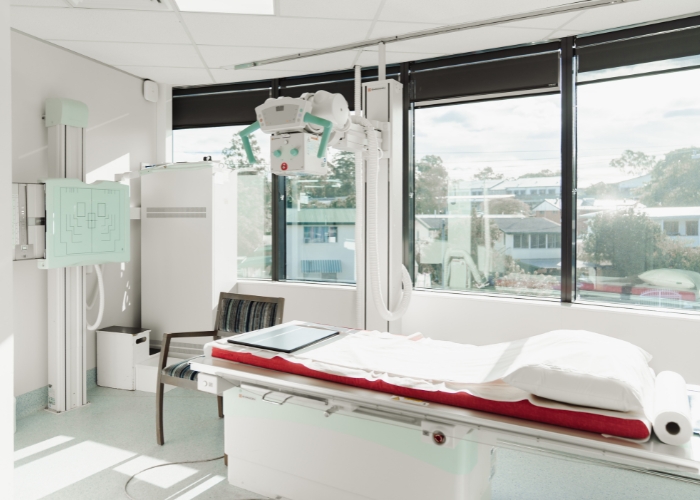
Fluoroscopy
At South Coast Radiology our Fluoroscopy services help diagnose and guide a wide range of medical procedures.
Fluoroscopy uses advanced low-dose technology, allowing our radiologists to see anatomy function while the body moves, demonstrating how organs, joints and soft tissues function in real time.
Fluoroscopy examinations include gastrointestinal studies, joint injections, spinal procedures and other diagnostic or therapeutic exams. This imaging technique ensures precision and safety at every step.

What is Fluoroscopy?
Fluoroscopy is a type of medical imaging that uses continuous X-rays to create a real time video of what’s happening inside your body.
Unlike a standard X-ray (which produces a single still image) fluoroscopy uses continuous x-ray to visualise ongoing movement, including swallowing, joint motion or the flow of contrast dye through organs and vessels.
It is also used to assist radiologists and specialists in performing targeted procedures with high accuracy and minimal discomfort.
Why might I need Fluoroscopy?
Fluoroscopy provides critical information for diagnosis or treatment.
Your doctor may refer you for a fluoroscopy examination to:
- Examine the digestive system (e.g. barium swallow, meal or follow through)
- Evaluate the bladder or urinary tract
- Guide spinal or joint injections
- Perform pain management procedures
- Assist with catheter placements or drain insertions
- Visualise swallowing or motility function
About Your Test

Before your appointment
Our staff will provide specific preparation instructions before your appointment. Preparation will depend on the type of fluoroscopic study you’re having:
- Some tests require fasting for 4–6 hours before your appointment.
- You may be asked to change into a gown and remove jewellery or metal items.
- If contrast dye is used, please let staff know if you have any allergies, diabetes, asthma or kidney problems.
- Bring your referral, Medicare card and previous imaging results for comparison.

On the day
Our experienced staff will ensure your comfort and safety throughout the process.
- You’ll be positioned on an examination table beneath the fluoroscopy unit.
- You may be asked to hold your breath or change position.
- Depending on the type of test a contrast agent (such as barium or iodine) it may be swallowed, injected or administered via catheter to highlight specific areas.
- The radiologist or radiographer will observe your body’s motion on a monitor in real time and take images as needed.
- Most fluoroscopy studies take 15–60 minutes to complete.

After your appointment
- You can usually return to normal activities right away.
- If you received oral or injected contrast, drink plenty of fluids over the next 24 hours to help flush it from your system.
- Your images will be reviewed by a specialist radiologist, who will send a detailed report to your referring doctor.
- Your doctor will then discuss the results and next steps with you.
Frequently Asked Questions
Modern equipment uses low-dose radiation and all exams are performed under strict safety standards.
The benefits of accurate diagnosis far outweigh the minimal radiation exposure.
Please tell staff if you are pregnant or suspect you may be pregnant.
Some procedures use contrast to make internal structures more visible. Staff will explain this beforehand and answer any questions you may have.
Fluoroscopy is generally painless. However, some patients may experience mild discomfort depending on the test type (for example, during an injection).
Yes. A referral from your doctor or specialist is required for Medicare rebates and to ensure the correct study is performed.
Get real-time insights into your body
Book your appointment at South Coast Radiology for precise fluoroscopic imaging.