Our services
Echocardiography
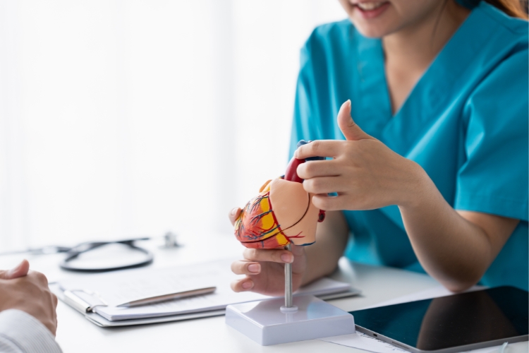
At South Coast Radiology, we provide advanced Echocardiography (also known as an Echocardiogram or Echo) to assess the structure and function of your heart in real time without radiation or discomfort.
This safe, non-invasive test uses ultrasound waves to create moving images of your heart, helping doctors evaluate how well it pumps blood and how the valves are working.
Performed by our highly trained cardiac sonographers and interpreted by specialist radiologists, your echocardiogram results provide crucial insight into your heart health.
What is an Echocardiogram?
An Echocardiogram is a diagnostic ultrasound of the heart.
It uses sound waves (not X-rays) to produce detailed, moving images of the heart’s chambers, walls, and valves, as well as blood flow patterns.
This test helps your doctor understand how efficiently your heart is functioning and detect a wide range of cardiac conditions such as:
- Heart valve disease
- Cardiomyopathy (thickened or weakened heart muscle)
- Congenital heart defects
- Pericardial Effusion (Fluid around the heart)
- Heart failure
- Reduced pumping function
Why might I need an Echocardiogram?
An echocardiogram is a key tool for understanding your overall heart health and guiding your treatment plan.
Your doctor may refer you for an Echocardiogram to:
- Investigate chest pain, palpitations, breathlessness or fatigue
- Assess heart murmurs or irregular rhythms
- Monitor known heart disease or heart valve conditions
- Evaluate the effects of certain medications or treatments
- Check heart function following surgery, chemotherapy or radiotherapy
About Your Test

Before your appointment
Our friendly cardiac team will guide you through the process and answer any questions before your scan begins.
- No fasting or special preparation is required.
- Continue taking your usual medications unless advised otherwise.
- Wear comfortable, two-piece clothing that allows easy access to your chest area for the echocardiogram.
- Bring your referral, Medicare card, and previous cardiac imaging results if available.

On the day
Our sonographers work with great care to ensure clear images and a comfortable experience.
- You’ll be asked to lie on an examination bed, usually on your left side.
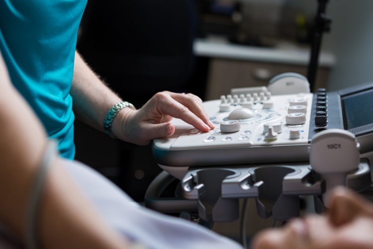
- A small handheld probe (called a transducer) with warm ultrasound gel is placed on your chest.
- The sonographer moves the probe to capture images of your heart in motion, which appear instantly on a monitor.
- You may hear “whooshing” sounds as the blood flow through your heart is measured.
- The test is painless and generally takes 30 minutes.

After your appointment
- You can return to normal activities immediately after your appointment.
- One of our specialist radiologists or cardiologists will review your images and send a comprehensive report to your referring doctor.
- Your doctor will then discuss the results and any recommended follow-up.
- If urgent findings are detected, results are prioritised and sent promptly to your healthcare provider.
Frequently Asked Questions
Yes. It’s a completely safe, non-invasive test that uses sound waves (not radiation).
No. You may feel light pressure from the probe on your chest, but there is no pain involved.
Yes. Ultrasound is safe during pregnancy.
Your images and report are provided through a secure online system to your doctor or healthcare provider within 24-48 hours.
Gain a clear understanding of your heart health
An Echocardiogram at South Coast Radiology can provide vital insights into your health.