Discover exceptional diagnostic imaging services at South Coast Radiology.
Our comprehensive offerings include x-ray, ultrasound, MRI, CT, dental imaging, nuclear medicine, breast imaging, bone densitometry, and interventional procedures.
Our patients and referrers trust us for high-quality imaging, precise reporting, and compassionate care.
Explore our services today for accurate diagnoses and superior patient care.
Bone Mineral Densitometry
A Bone Mineral Densitometry scan is a quick and painless procedure that accurately measures bone strength and density.
These scans help diagnose osteoporosis and assess fracture risks, guiding proactive measures for better bone health.
It provides information about the possible fracture risk to your bones.
A stereotactic breast biopsy is a specialised imaging-guided procedure used to obtain tiny samples of breast tissue for examination under a microscope.
The term “stereotactic” refers to the 3D guidance system used — multiple low-dose X-ray images are taken from different angles, and computer technology pinpoints the exact location of the abnormality within the breast.
A Calcium Score scan is a specialised CT scan that detects and measures calcium deposits in the walls of your heart’s arteries.
Calcium build-up can indicate atherosclerosis, a condition where plaque hardens and narrows the arteries, potentially leading to heart attack or stroke.
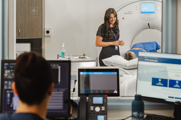
A CT (computed tomography) scan combines X-rays with advanced computer processing to create highly detailed 3D images of your body.
Unlike a standard X-ray that shows one image, a CT scan takes many thin “slices” from different angles giving your doctor a comprehensive view of what’s happening inside your body.
CT scans are commonly used to examine the chest, abdomen, pelvis, head, spine, and joints, and play an important role in diagnosing disease, injury, or internal abnormalities.
Dental Imaging uses advanced X-ray technology to capture detailed images of the teeth, jaws, and facial structures, supporting accurate diagnosis and treatment planning.
At South Coast Radiology we offer the following dental imaging services:
Fluoroscopy is a type of medical imaging that uses continuous X-rays to create a real time video of what’s happening inside your body.
Unlike a standard X-ray (which produces a single still image) fluoroscopy uses continuous x-ray to visualise ongoing movement, including swallowing, joint motion or the flow of contrast dye through organs and vessels.
Interventional Radiology uses advanced imaging to guide fine instruments such as needles, catheters, or electrodes directly to the area of concern.
These minimally invasive procedures allow our radiologists to diagnose or treat conditions through tiny skin punctures, helping to manage pain in the joints, spine, soft tissues, and tendons – often providing lasting relief without the need for surgery.
In collaboration with the National Lung Cancer Screening Program (NLCSP), we have partnered with Chest Scan to provide low-dose chest CT to catch lung cancer in its early stages.
A low-dose chest CT (LDCT) is a specialised imaging test with minimal radiation exposure to create detailed images of the lungs.
Lung cancer screening allows for us to assess the health of your lungs and monitor any changes over time, detecting all types of lung cancer at a stage when treatment is most effective.
Mammography is a specialised low-dose X-ray of the breast that produces detailed images of breast tissue from different angles.
It’s considered the gold standard for early breast cancer detection and is also used to monitor known breast conditions over time.
Our 3D digital tomosynthesis captures thin layers of breast tissue to create a more detailed view, improving cancer detection, reducing unnecessary call-backs and particularly beneficial for women with dense breast tissue.
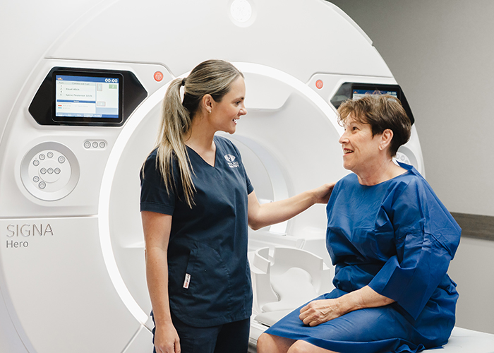
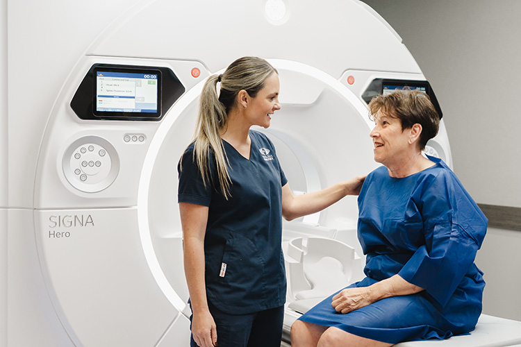
MRI (Magnetic Resonance Imaging) uses powerful magnets, radiofrequency pulses, and computer technology to create detailed cross-sectional or 3D images of your body’s internal structures.
The resulting signals are processed to reveal subtle differences in tissues, making MRI highly effective for identifying inflammation, nerve compression, soft-tissue tears, and other internal abnormalities not visible on X-ray or CT.
At South Coast Radiology we provide a range of MRI imaging, including:
Nuclear Medicine is a highly specialised branch of medical imaging that enables us to visualise organ and tissue function in real time.
Rather than imaging anatomy alone (as with CT or MRI), Nuclear Medicine reveals the physiology i.e. how well parts of your body are working.
To perform these scans, a small dose of a radiopharmaceutical (a tracer) is introduced into your body, usually by injection, inhalation, or oral intake.
The tracer accumulates in the target organs or tissues. As it emits gamma rays, a gamma camera (or PET scanner) detects these emissions and constructs images showing how the tracer is distributed.
An Obstetric Ultrasound uses sound waves (not radiation) to produce real-time images of your developing baby and uterus.
This scan is completely safe and painless, and provides valuable information about your baby’s size, growth, anatomy and position as well as your placenta and amniotic fluid levels.
At South Coast Radiology we offer a range of Obstetric Ultrasounds including:
OncoBeta® (Rhenium-SCT®) is a non-invasive therapy that uses the radioactive isotope Rhenium-188 to precisely deliver beta radiation to the affected area of the skin.
The radiation penetrates only the top layers of the skin, destroying cancer cells while sparing healthy tissue underneath. The treatment is painless, quick, and performed as an outpatient procedure within our Nuclear Medicine department.
This therapy is especially effective for patients with:
PET/CT combines two powerful imaging techniques, Positron Emission Tomography (PET) and Computed Tomography (CT), into a single scan that provides both functional and anatomical information.
This highly specialised test helps doctors detect cancer, monitor treatment response, and assess organ function with remarkable precision all in one visit.
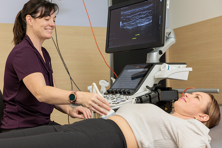
Ultrasound works by sending harmless sound waves into the body. These waves bounce back as echoes and are converted into live images by a computer.
Because ultrasound provides real-time imaging, it can show movement — such as a beating heart, flowing blood, or fetal motion — not just static pictures.
It’s commonly used to assess:
Abdominal and pelvic organs (liver, gallbladder, kidneys, uterus, prostate)
An X-ray uses a small amount of ionising radiation to produce images of the body’s internal structures. Denser tissue, like bone, appears white, while softer tissue appears in shades of grey.
Modern digital detectors make the process fast, efficient, and are lower in radiation than ever before.