Our services
Nuclear Medicine
At South Coast Radiology, our Nuclear Medicine service provides powerful insight into how organs and tissues function, not just how they look.
By using tiny amounts of radioactive tracers (radiopharmaceuticals), we can detect abnormalities in your body’s major systems earlier and more accurately than many other modalities.
Recognised as the Gold Coast’s leading Nuclear Medicine and PET/CT provider, we deliver precision diagnostics backed by compassionate care.
For eligible patients, many of our scans are bulk billed under Medicare.
What is Nuclear Medicine?
Nuclear Medicine is a highly specialised branch of medical imaging that enables us to visualise organ and tissue function in real time.
Rather than imaging anatomy alone (as with CT or MRI), Nuclear Medicine reveals the physiology i.e. how well parts of your body are working.
To perform these scans, a small dose of a radiopharmaceutical (a tracer) is introduced into your body — usually by injection, inhalation, or oral intake.
The tracer accumulates in the target organs or tissues. As it emits gamma rays, a gamma camera (or PET scanner) detects these emissions and constructs images showing how the tracer is distributed.
Areas of abnormal uptake can indicate disease, dysfunction, or pathology.
This capability makes Nuclear Medicine uniquely useful for diagnosing conditions before structural changes appear on conventional imaging.
Common Nuclear Medicine (and PET) Scans
Each protocol is tailored to your referral and the clinical question at hand.
Some of the most frequently performed studies we perform include:
-
Bone scans
Evaluate bone pain, fractures, metastases, arthritis or infections
-
Thyroid scans
Assess function, nodules, or hyperthyroidism
-
Renal scans
Examine kidney function, drainage and perfusion
-
Lung scans (V/Q scans)
Detect pulmonary embolism or assess lung ventilation/perfusion
-
Cardiac/Myocardial Perfusion Scans
Assess blood flow to heart muscle, detect ischemia
-
Gastrointestinal and gastric emptying scans
Evaluate swallowing, reflux, bleeding or motility
-
OncoBeta® Skin Cancer Therapy
Treatment using radionuclide technology for selected skin lesions
-
PET/CT
Combines functional and anatomical imaging for oncologic, neurologic and cardiac applications
Why might I need a nuclear medicine test?
In many cases, Nuclear Medicine findings are complementary to CT, MRI or ultrasound and can provide crucial additional information.
Your doctor or specialist may request a Nuclear Medicine study to:
- Assess how well an organ is functioning (rather than just its appearance)
- Detect disease earlier, even before structural changes become apparent
- Monitor disease progression or response to therapy
- Guide treatment plans for cancer, cardiac disease, renal disease, or endocrine disorders
- Localise sources of pain, infection or abnormal biochemical markers
About Your Test

Before your appointment
- A referral from your GP or specialist is required.
- While booking, our team will provide you with specific preparation instructions depending on the scan you require e.g. fasting, hydration, or (in some cases) temporarily stopping certain medications.
- Let us know if you are pregnant, breastfeeding, diabetic, or have allergies to medications or contrast agents.
- Wear comfortable, loose clothing (remove jewellery or metallic objects in the area to be imaged).
- Bring your Medicare card, referral, and any prior imaging or reports to aid comparison.
- At the appointment, you may be asked to wear a gown and leave personal items (wallet, phone) outside the imaging room to reduce interference.

On the day
- The tracer is administered (via injection, inhalation, or oral route), after which there may be a waiting period to allow it to accumulate in the target area.
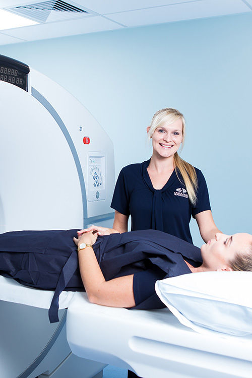
- You’ll be positioned under the gamma or PET camera. The camera moves to image the region(s) of interest while you lie still.
- You may be asked to change position or hold your breath briefly, depending on the study.
- The scan is painless and non-invasive. Most patients report no discomfort.
- The duration of the exam varies depending on the type. Typically these tests last from 30 minutes up to 2 hours or more in complex examinations.
- Throughout the scan, our technologists monitor you and ensure image quality and patient safety.

After your appointment
- You may resume most activities immediately after the scan.
- Drink plenty of fluids (unless otherwise advised) over the next day to help clear tracer from your body.
- The radiopharmaceutical decays or is eliminated naturally and poses no ongoing risk.
- Images and a detailed interpretive report are prepared by our specialist Nuclear Medicine radiologists and sent to your referring doctor via secure channels.
- In many cases, you will also receive (or have access to) a printed or electronic copy to take to your next consultation.
Frequently Asked Questions
You’ll receive specific instructions at booking. Depending on the type of tst this could include fasting, avoiding caffeine, or skipping certain medications.
The risks are minimal. Radiopharmaceutical doses are small and tailored to be safe for diagnostic use. Adverse reactions are exceedingly rare.
A tracer is introduced, you wait as it distributes, then images are captured using a gamma or PET camera. The process is painless.
Time depends on the type of scan: simple scans may finish within 30–45 minutes; more complex or multi-phase studies may take 1–2 hours or more.
For Medicare-eligible studies, Nuclear Medicine scans are bulk billed. For non-eligible patients, your booking team will advise you of costs at the time of booking.
Risks & Safety
The dose of radiation used is very low and carefully calculated to minimise risk while giving diagnostic value.
Side effects or adverse reactions are extremely rare, as tracers are rigorously tested and chosen for safety.
Because the tracers are short-lived, any residual radioactivity leaves your body quickly.
If you are pregnant or breastfeeding, a Nuclear Medicine scan may be delayed or replaced with an alternate modality unless clinically critical.
With kidney or other organ impairments, tracer selection and dosing may be adjusted for safety.