
South Coast Radiology (SCR) is expanding its specialised PET/CT imaging services, with FDOPA PET/CT brain scans now available to help specialists assess Parkinson’s disease and other movement disorders.
This advanced imaging technique provides valuable information about dopamine activity in the brain, helping doctors investigate symptoms and support clinical decision-making when a diagnosis is uncertain.
What is an FDOPA PET/CT brain scan?
FDOPA PET/CT is a specialised nuclear medicine imaging procedure that uses a small amount of a radioactive tracer called fluorine-18 fluorodopa (¹⁸F-FDOPA).
The tracer helps visualise aspects of dopamine production and processing in the brain. Dopamine is a chemical messenger involved in controlling movement, and changes in the brain’s dopaminergic system are associated with Parkinson’s disease.
By showing patterns of tracer uptake in specific areas of the brain, an FDOPA PET/CT scan can provide additional information to help specialists assess Parkinsonian symptoms and distinguish between possible causes.
How can FDOPA PET/CT help?
Diagnosing Parkinson’s disease can sometimes be challenging, particularly when symptoms overlap with other neurological conditions.
An FDOPA PET/CT scan can provide complementary information alongside a patient’s medical history, clinical examination and other investigations. This may help a treating specialist clarify a diagnosis and guide decisions about ongoing care.
The scan is not required for everyone with suspected Parkinson’s disease. A specialist will determine whether it is appropriate based on the individual’s symptoms and clinical circumstances.
What can patients expect during the scan?
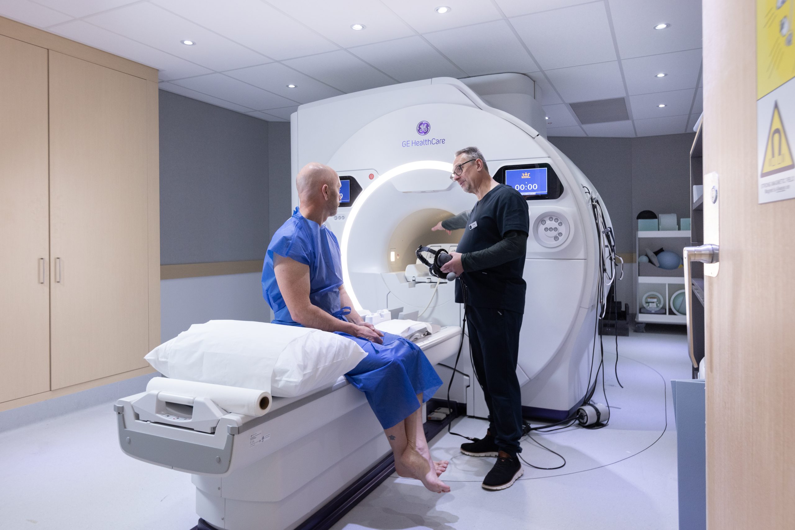
The procedure involves an injection of the FDOPA tracer, followed by a period of rest while it distributes through the body. The patient then lies on the PET/CT scanner bed while images are acquired.
Patients should allow approximately 2.5 to 3 hours for the appointment, although timing can vary. Specific preparation instructions will be provided before the scan, and it is important to follow these carefully.
The PET/CT images combine information about tracer activity with detailed anatomical images, allowing the specialist to assess the distribution of the tracer within the brain.
Access FDOPA PET/CT at South Coast Radiology
FDOPA PET/CT brain scans are now available through South Coast Radiology’s two specialty PET/CT clinics (Southport and Tugun), providing patients and referring specialists access to this specialised imaging option to support the assessment of Parkinson’s disease.
If you or someone you care for is experiencing symptoms that may be associated with Parkinson’s disease, speak with your treating doctor or neurologist about the most appropriate investigations.
For referring practitioners:
Please contact South Coast Radiology to discuss FDOPA PET/CT referrals, appointment availability, preparation requirements and applicable fees.
South Coast Radiology is committed to providing access to specialised diagnostic imaging to support informed clinical decisions and patient care.