
Our services
3D Pregnancy Ultrasound
At The Women’s Imaging Centre, we understand how special pregnancy is and we’re honoured to be part of your journey.
Our 3D Pregnancy Ultrasound provides useful information about the uterus, ovaries and baby to enable appropriate antenatal care. Fetal size and heart rate are checked at all stages of pregnancy, as well as the maternal uterus, ovaries and cervix.
Using advanced, low-intensity ultrasound technology, our skilled sonographers create a safe and caring environment to capture clear, high-resolution images.
What is a 3D Pregnancy Ultrasound?
Ultrasound provides useful information about the uterus, ovaries and baby that enables appropriate antenatal care.
Pregnancy ultrasound is a non-invasive painless examination to assess fetal growth, position and anatomy, and placental site, as well as the maternal uterus and ovaries.
Ultrasound can detect many, but not all abnormalities. Demonstrating a fetal abnormality on ultrasound depends on many factors, including fetal age and position at the time of the ultrasound, and the size and type of abnormality.
There is very little preparation required for these exams. You should have some fluid in the bladder, and it is recommended to wear clothing that allows easy access to the entire abdominal region.
Medicare provides a rebate for ultrasound during the first and second trimester, and when there is a clinical concern.
Types of Pregnancy Ultrasounds
At The Women’s Imaging Centre, we perform a range of pregnancy scans, including:
-
Early Pregnancy/Dating Scan
Around 6-10 weeks
Confirms pregnancy, gestational age, heartbeat and location. -
CFTS Nuchal Translucency Scan
First Trimester: 11-14 weeks
Assesses early fetal development and may include optional genetic screening (at your doctor's request). -
Morphology Scan
Second Trimester: 18-22 weeks
A detailed structural assessment of your baby’s organs, limbs, spine and facial features, and checks placenta and amniotic fluid. -
Growth and Wellbeing Scan
Third Trimester: From 28 weeks onward
Monitor your baby’s position, growth, and overall health in later stages of pregnancy. -
Doppler Blood Flow Studies
Assess blood flow to the placenta and baby for high-risk or complex pregnancies.
About Your Test

Before your appointment
- For early pregnancy scans, you may be asked to attend with a comfortably full bladder (drink water 1 hour before your appointment).
- Later-term scans usually don’t require bladder preparation.
- Wear a two-piece outfit for comfort and convenience.
- Please bring your referral, Medicare card, and any previous imaging if applicable.
- Let our staff know if you’re having twins or have a high-risk pregnancy so extra time can be allocated.
- Our sonographers take every care to make you comfortable and answer your questions throughout.

On the day
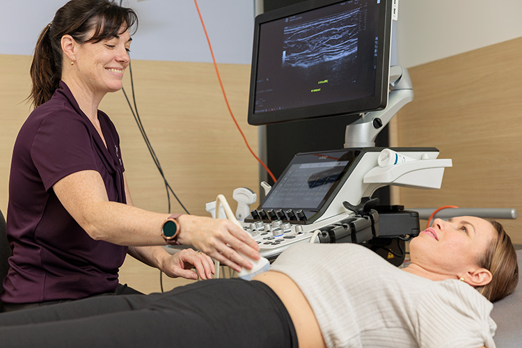
- You’ll lie on a padded examination bed while the sonographer applies a small amount of warm gel to your abdomen.
- A handheld probe (transducer) is moved gently across your skin to capture live images.
- You’ll be able to view your baby on the screen during the scan. Many parents find this a wonderful bonding moment.
- Occasionally, for early pregnancy or detailed imaging, a transvaginal ultrasound may be recommended; this is safe and will be explained beforehand.
- Most scans take 20–60 minutes, depending on your stage of pregnancy.

After your appointment
- You can return to normal activities immediately.
- Your images and a detailed report are reviewed by a specialist radiologist and sent directly to your referring doctor or midwife.
- If anything requires follow-up, your healthcare provider will discuss this with you.
- Some of our clinics can also provide printed images or digital access to share with family.
Frequently Asked Questions
Yes. Ultrasound has been used in pregnancy for decades and has no known risks to mother or baby.
Usually between 6–10 weeks to confirm the pregnancy and heartbeat.
Yes, in most cases a partner or family member may attend. Please check at the time of booking.
Yes, the morphology scan (around 20 weeks) can usually reveal the baby’s sex if desired.
Yes. A valid referral from your GP, specialist, or midwife is required for Medicare rebate eligibility.
Safety & Comfort
Obstetric ultrasound is completely safe — it uses sound waves, not radiation.
All scans are performed by accredited sonographers trained in obstetric imaging.
Our team strives to create a calm and positive experience for every expectant parent.
Partner attendance is welcome, and we’ll do our best to accommodate your support person.
For your comfort, we recommend that babies and young children do not attend your appointment, as obstetric ultrasound scans can take up to 60 minutes or longer to complete.
Enjoy peace of mind
At The Women’s Imaging Centre you’ll experience gentle, expert obstetric care performed by our leading sonographers and radiologists.