Our services
3D Mammography
At The Women’s Imaging Centre, we’re dedicated to supporting breast health through advanced 3D digital mammography and comprehensive breast imaging services.
Mammography is a quick, safe, and highly accurate screening tool that helps detect early changes in breast tissue, often before they can be felt. This allows for timely diagnosis and the best chance of successful treatment.
Our clinics feature state-of-the-art 3D tomosynthesis (3D mammography) systems that deliver superior image quality with minimal radiation exposure.
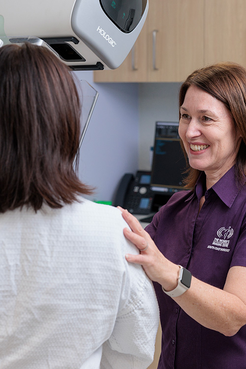
Every mammogram is performed by our compassionate, female radiographers, accredited with the Australian Institute of Radiography (AIR) and experienced in advanced breast imaging techniques. They understand that every patient deserves a calm, respectful, and comfortable experience.
What is 3D Mammography?
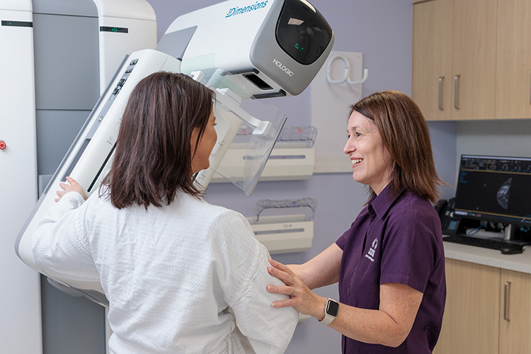
3D Mammography (Breast Tomosynthesis) is the latest, most advanced technology for early detection of Breast Cancer. The Women’s Imaging Centre performs 3D Mammography Breast Tomosynthesis in conjunction with a traditional 2D digital mammogram, and quite often a breast ultrasound.
Instead of viewing the complexities of the breast tissue in one flat 2D image, the radiologist can scroll through the images of your entire breast one layer at a time. A common analogy for 3D Mammography is likened to the pages in a book. Instead of just seeing the cover of the book, 3D mammography is like seeing all the information inside page by page from cover to cover. 3D mammography allows the radiologist to view the breast tissue in 1mm slices and eliminates the confusion of overlapping tissue.
Mammography is a low-dose X-ray used to assess breast disease in symptomatic women, and as a screening tool in the wider population. Mammography can show small cancers well before a lump is palpable, and early detection increases the overall effectiveness of cancer treatment.
Around 10% of all breast cancers will not be identified by mammography alone. Ultrasound is often used to further assess breast tissue and improve detection rates.
At the Women's Imaging Centre we offer both:
-
Screening Mammography
Aimed at early detection for women with no symptoms.
-
Diagnostic Mammography
For women experiencing symptoms or who require further investigation after a screening mammogram.
Understanding Breast Density
Breast density refers to the amount of glandular and fibrous tissue compared to fatty tissue in your breasts.
Dense breasts are common but can make it harder to detect changes on a mammogram.
If you have dense breast tissue, your doctor may recommend additional imaging such as breast ultrasound or MRI to ensure every area is clearly assessed.
Why might I need a Mammogram?
Your doctor may refer you for mammography to:
- Screen for early signs of breast cancer
- Investigate lumps, pain, nipple discharge, or skin changes
- Monitor previously detected abnormalities
- Assess post-surgical changes, scar tissue, or implants
- Evaluate findings seen on other breast imaging tests
Regular mammograms can detect changes years before they are felt or visible, improving outcomes and reducing the need for more invasive treatment.
About Your Test

Before your appointment
Our friendly staff will guide you through every step and ensure you know what to expect before your scan.
- Try to schedule your mammogram one week after your menstrual period, when breasts are less tender.
- Avoid deodorant, perfume, powder, or lotion on the day of your scan, as these can appear as artefacts on images.
- Wear a two-piece outfit for easier removal of clothing from the waist up.
- Bring previous breast imaging for comparison, if available.
- Inform our team if you have breast implants, are pregnant, or breastfeeding.

On the day
You’ll stand in front of the mammography unit while one breast at a time is gently positioned between two compression plates.
- Compression lasts only a few seconds but it’s essential for clear, accurate images while minimising radiation dose.
- The technologist will capture views of each breast – from the top and from the side.
- Occasionally additional images are required at the radiologist’s discretion.
- The entire procedure takes about 15 minutes.
- If any additional images are needed for clarification, our team will let you know before you leave.

After your appointment
- You can return to normal activities immediately.
- Your images will be reviewed by two specialist breast radiologists, and a detailed report will be sent directly to your referring doctor.
- If additional imaging (such as ultrasound or MRI) is recommended, our team will coordinate this promptly to avoid delay in results.
- Results are usually available to your referring doctor within 24–48 hours.
Frequently Asked Questions
Most women aged 40 and over should have a screening mammogram every one to two years, depending on personal and family history.
Yes. Our radiographers use special techniques and additional images to safely capture clear images around implants.
You can still have mammograms. Just let your radiographer know about your surgery so positioning can be adjusted appropriately.
Yes. A referral from your GP or specialist ensures the correct test is performed.
Mammography is generally avoided during pregnancy unless absolutely necessary, but your doctor will discuss the safest options with you.
Safety and Comfort
Mammography uses very low-dose X-rays, making it extremely safe.
The compression may feel uncomfortable for a few seconds but should not be painful.
All equipment meets the highest Australian radiation safety standards.
Our experienced female radiographers are dedicated to performing each scan gently, efficiently, and with respect.
Experience compassionate care
Protect your breast health with our expert imaging.
Contact your nearest South Coast Radiology clinic or speak to your doctor about booking your mammogram today.